Problem with lung motion in radiation therapy and current 4D methods
5DCT brings accurate tumor delineation and treatment planning
Patients with inconsistent breathing patterns complicate 4DCT acquisition. Even with coaching, some patients may fail to produce regular breathing traces, increasing the likelihood of motion-related inaccuracies in imaging and treatment delivery.
Our 5D solution – Meet Respirati 5DCT®
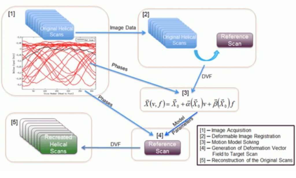
We start with a series of conventional helical CT scans while monitoring breathing patterns, that are generally irregular. This provides us with multiple breath-hold-like images sampled over two minutes (to sample multiple breathing cycles) that are compared with each other (using deformable image registration) to characterize the tumor motion and correlating it with the breathing amplitude and rate. We combine this tumor motion information (x, y, and z), with the breathing information (amplitude and rate) to generate artifact-free images for treatment planning. Respirati 5DCT has been shown to outperform all commercially available image reconstructions methods to prepares the radiation oncology team with the most accurate tumor and motion margin (ITV) delineation for treatment planning.
The 5DCT name stems from the motion model’s description of tissue displacement as a function of the five variables: x, y, and z position, breathing amplitude, and breathing rate.


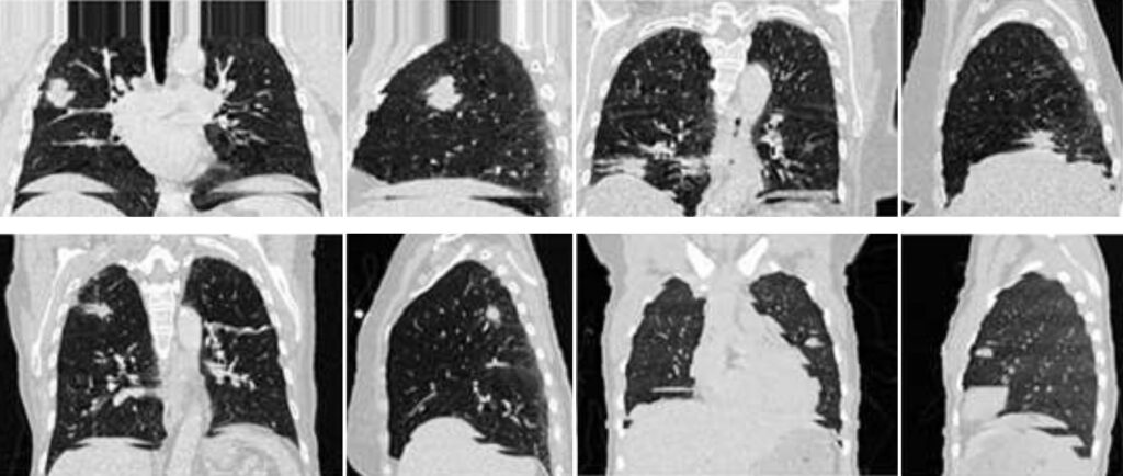
4DCT (before)
Limitations of 4DCT methods in radiation therapy
Despite being the most widely used technology for reconstructing lung tumor motion, 4DCT methods do not accurately measure tumor volume and breathing motion. The main reason is that most patients breathe irregularly, leading to erroneous sampling and subsequent image artifacts.
As a result, 4DCT images often misrepresent tumor volumes.

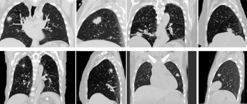
5DCT®
Respirati 5DCT® combines a novel CT scanning technique and a motion model to provide artifact-free images and accurate tumor volume and motion for patients who breathe irregularly.