The Evolution of 4DCT in Lung Cancer Radiation Therapy
Before 4DCT, standard imaging techniques such as free-breathing CT or breath-hold CT were used. However, these approaches often failed to account for the full extent of tumor motion during the respiratory cycle, leading to uncertainties in treatment planning. The early days of four-dimensional computed tomography (4DCT) for imaging lung cancer patients in radiation therapy were marked by the need to address respiratory motion and improve tumor localization. The development of 4DCT provided a breakthrough by offering breathing phase-specific images that allowed clinicians to visualize and quantify tumor motion.
Early Development and Challenges
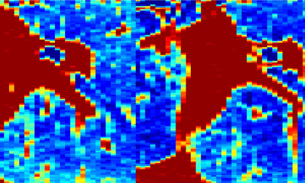
Early 4DCT systems relied on phase-based binning, in which images were retrospectively sorted into different phases of the breathing cycle. This method aimed to reconstruct motion-resolved CT datasets, capturing how tumors moved over time. Early studies revealed that image artifacts caused by irregular breathing could impact the accuracy of tumor motion estimations. Later, amplitude-based sorting was developed, often improving upon phase-based gating, but irregular breathing still caused some amplitudes to lack data, leading to image artifacts. Despite these challenges, 4DCT quickly demonstrated significant advantages over static CT by allowing the definition of internal target volumes (ITV), enabling better tumor coverage and reduced normal lung dose.
Integration into Image-Guided Radiation Therapy (IGRT)
- As the use of 4DCT expanded, its role in IGRT became evident.
- Early adopters explored ways to integrate 4DCT into IGRT workflows, where tumors were imaged on the treatment table and ITV margins were evaluated.
- Studies comparing pre-treatment 4DCT scans with daily imaging using 4D cone-beam CT (4D-CBCT) confirmed that tumors could shift significantly between fractions.
- This reinforced the need for motion management strategies, such as on-table imaging, to validate or modify margins.
- These findings drove the development of respiratory-gated radiation delivery, where radiation beams were turned on only during specific phases of the breathing cycle to minimize exposure to surrounding healthy tissues.
Standardization and Advancements
- By the late 2000s, 4DCT had become a standard imaging modality for lung cancer radiation therapy planning.
- 4DCT influenced the development of stereotactic body radiation therapy (SBRT) protocols.
- Research explored dose accumulation and interplay effects, particularly in intensity-modulated radiation therapy (IMRT) and volumetric-modulated arc therapy (VMAT).
- The early years of 4DCT laid the foundation for modern motion management strategies.
- Accurate tumor motion assessment remains essential for optimizing radiation dose delivery while minimizing toxicity to normal lung tissue.
Ironically, the fundamental need for 4DCT—measuring and characterizing breathing motion—was greatest for patients with irregular breathing. However, 4DCT was least reliable for these patients. More than 20 years later, this limitation continues to highlight the need for a 4DCT replacement.
Daniel Low, PhD
Washington University Saint Louis
